Technology
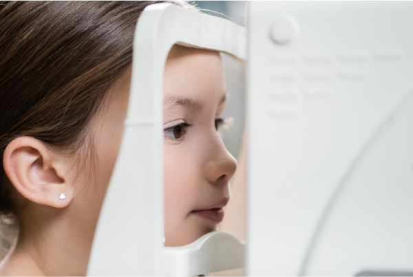
QUAD-IMAGING SMART MIRROR
Take all the guesswork out of getting your new glasses! See yourself in the eyewear of your choice in our exclusive Quad-Imaging smart mirror system. Patients can view four images of themselves on a monitor to compare the ‘before look’ with a new look’ side by side. No more squinting at handheld mirrors is trying to see. Quad-Imaging is the easiest and smartest part of your eyewear makeover.

CONFOCAL SCANNING LASER OPHTHALMOSCOPY (SLO)
Eye Care & Wear utilizes the HEIDELBERG SPECTRALIS ultra-high-speed, high-resolution SLO retina scanner for retinal imaging and analysis for early detection of glaucoma, macular degeneration, and other diseases of the retina such as diabetic and hypertensive retinopathy. The SPECTRALIS is based on next-generation Fourier-Domain Optical Coherence technology and allows Dr. Rustagi to visualize retinal tissue with ultra-high clarity in a fraction of seconds.
A laser light scans the retina in 24-millisecond sequential scans, starting above the retinal surface, then capturing parallel images at increasing depths. The stacks of images can be combined to create a three-dimensional (3-D) topographic image of the retina. Images are aligned and compared using TruTrack™ software for both individual examinations and for detecting change between examinations. The same technology is used in the SPECTRALIS Retina Module.
SLIT LAMP VIDEO CAMERA
Dr. Rustagi utilizes the REESEVIT Digital Video Camera to photograph the exterior or interior surface of your eye, including the cornea, retina, optic disc, macula, and posterior pole (i.e., fundus). He uses these images or videos to then help diagnose and monitor the progression of diseases such as glaucoma, dry eyes, pink eye, and many more. By using this advanced technology, we are able to photo document and monitor small changes to your eyes over time, which is essential for accurate detection and proper management of the disease.
SPECULAR MICROSCOPE
New to our office is our T3000 Specular Microscope. With this corneal imaging system, Dr. Rustagi can now assess multiple potential issues with your corneal health long before they can do permanent damage. Diseases such as dry eyes, genetic and age-related disorders can now be diagnosed and treated. If left undiagnosed, the normally clear cornea can become clouded, thus decreasing vision. Patients that over wear and sleep in their contacts are excellent candidates for early detection of any problems which may occur.
RetEval ERG- Diabetic Module
One of the most pressing concerns in primary eye care is accurately predicting which diabetic retinopathy patient is going to progress. Structural health assessed through imaging is only half of it. Functional diagnostics are vital.
We use a state of the art, portable and non-invasive RETeval® Device. It combines functional retina cell stress testing with pupil light response. This powerful combination provides a superior risk assessment for which patients are going to require an ocular intervention, compared to the current Standard of Care. Patients who have scored greater than 23.4 have an 11 times greater risk of progressing in the next 3 years.
Clinical management of diabetic retinopathy is improved by adding electroretinography to fundus photographic information in assessing the risk of the need for intervention.
The RETeval device helps our doctors to:

Detect
Detect various retina and optic nerve diseases earlier.
Predict
Predict the progression of the disease
Follow
Then follow up on the course of disease
Monitor
Monitor treatment successNext generation OCT module
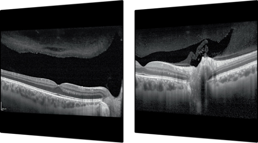
The SPECTRALIS® OCT2 Module is the next generation OCT technology for the SPECTRALIS platform, offering enhanced image quality and a faster scan speed. It improves clinical workflow and increases patient comfort by shortening examination times.
Optical Coherence Tomography (OCT) is a non-invasive imaging test that uses light waves to take cross-sectional pictures of your retina. With OCT, each of the retina’s distinctive layers can be seen with details discernible to micrometres.